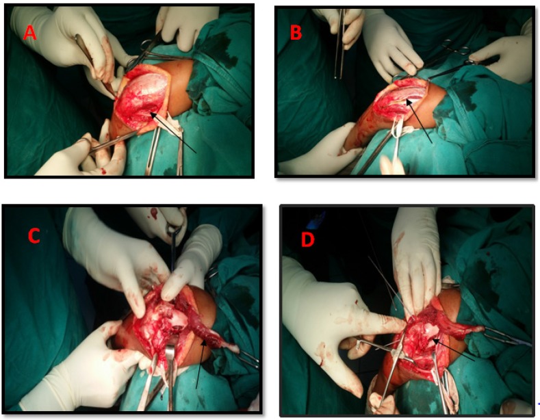
Fig. 1.
A: Skin incision and superficial dissection with isolation of ulnar nerve (black arrow); B: Inverted “V” shaped triceps tongue incision (black arrow); C: Intra-articular fracture distal humerus with triceps belly reflected (black arrow); D: Intra-articular fracture of distal humerus fixed with kirshner wires(black arrow

