FIG 5.
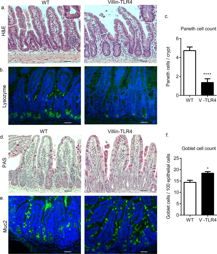
TLR4 regulates EC differentiation in the small intestine. (a) Histological images of ileum tissues from WT and villin-TLR4 littermate mice at 8 weeks of age. The inset shows a lower number of Paneth cells per crypt in villin-TLR4 mice than in WT mice. (b) Reduced lysozyme (green) staining in the ileum tissue of villin-TLR4 mice. (c) Graph of the number of Paneth cells (mean ± the standard error of the mean) in ileal sections of small intestine tissue as determined by H&E staining. Higher numbers of goblet cells in the ileum tissue of villin-TLR4 mice than in that of WT mice are indicated by periodic acid-Schiff (PAS) (d) and Muc2 (green) immunofluorescence (e) staining. (f) Graph of goblet cell numbers (mean ± the standard error of the mean) determined in PAS-positive cells in ileum tissue sections from WT and villin-TLR4 mice. For immunofluorescent images (b, e), nuclei were stained with DAPI (blue). Images are representative of four mice per group. Scale bars, 200 μm. Magnification, ×20. *, P < 0.05; ****, P < 0.0001 by Student's t test.

