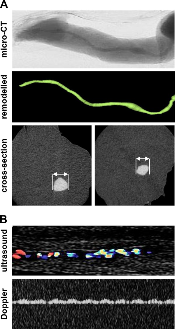
FIG 2.

Microcomputed tomography and Doppler ultrasound analysis of umbilical cord veins. (A) Umbilical cord veins were perfused with MicroFil contrast solution, recorded by microcomputed tomography, and digitally processed; total length analyzed, 1.8 cm; diameter, 1.3 (left) or 0.9 (right) mm. (B) For ultrasound analysis, umbilical cord veins were washed and infected with barium sulfate (10%, resuspended in PBS).
