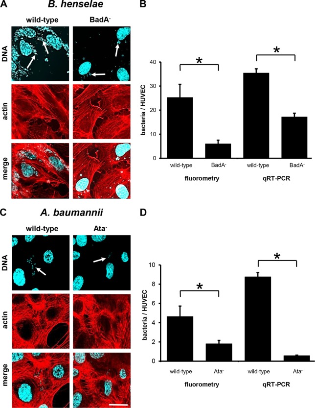
FIG 4.
TAA-dependent adhesion of B. henselae and A. baumannii to HUVECs (static infection). HUVECs (1.0 × 10⁵) were seeded on coverslips and infected with the respective bacteria (for B. henselae at an MOI of 100; for A. baumannii at an MOI of 200) for 60 min. For fluorescence microscopy (A, C), actin cytoskeleton of ECs was stained with TRITC-phalloidin (red), bacteria and nuclei were stained with DAPI (blue), and images were digitally overlaid (merge). Scale bar, 20 μm. (B, D) Adherence to ECs was determined in parallel by using fluorometry and qRT-PCRs. *, P < 0.05.

