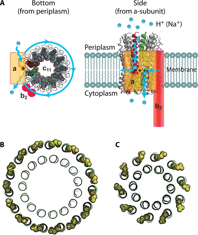
FIG 3.
Structure of Fo. (A) Crystal structure of the c11 ring of Na+-transporting Fo from Ilyobacter tartaricus (PDB code 1YCE). The blue spheres in the middle of the c11 ring represent bound Na+ ions. The stator ab2 complex is shown in the schematic drawing. The a subunit has 2 hemichannels, each open to the periplasmic space or the cytoplasmic space. A proton transferring between the a and c subunits accompanies the rotation of the c ring. Two c-subunit monomers at the interface of the a subunit are shown in red and green, respectively. (B) “Ion-locked” conformation of cGlu62 (yellow sphere representation) in the crystal structure of the H+-transporting c15 ring from Spirulina platensis (PDB code 2WIE). (C) “Ion-unlocked” conformation of cGlu59 (yellow sphere representation) in the crystal structure of the H+-transporting c10 ring from yeast mitochondria (PDB code 3U2F).

