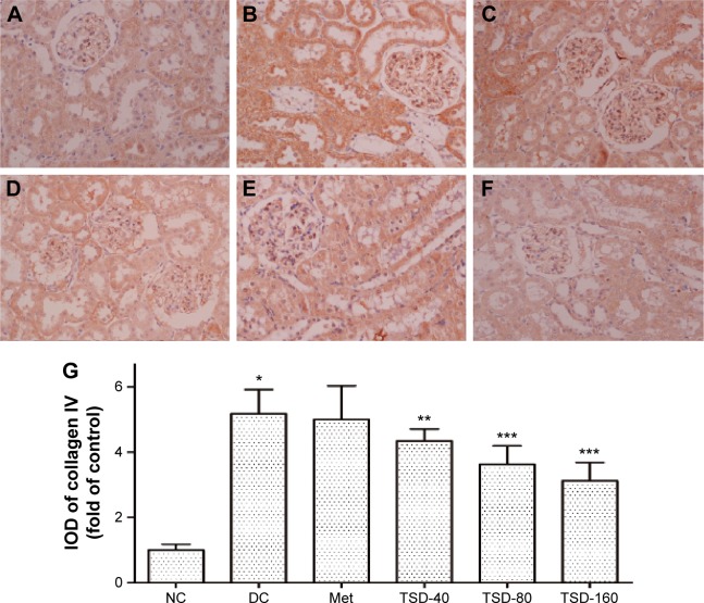
Figure 6.
Effect of TSD on renal immunohistochemical stain of collagen IV in diabetic rats.
Notes: The brown area was the positive expression area, enlarging 400× under light microscope. (A) NC group, (B) DC group, (C) Met group, (D) TSD-40 group, (E) TSD-80 group, (F) TSD-160 group, and (G) IOD of collagen IV. Values are presented as mean ± SD for eight rats in each group. *P<0.01 as compared with the normal control group. **P<0.05 as compared with the diabetic control group. ***P<0.01 as compared with the diabetic control group.
Abbreviations: IOD, integrated optical density; NC, normal control group; DC, diabetic control group; Met, metformin hydrochloride group; TSD, total saponin of Dioscorea hypoglaucae Palibin.

