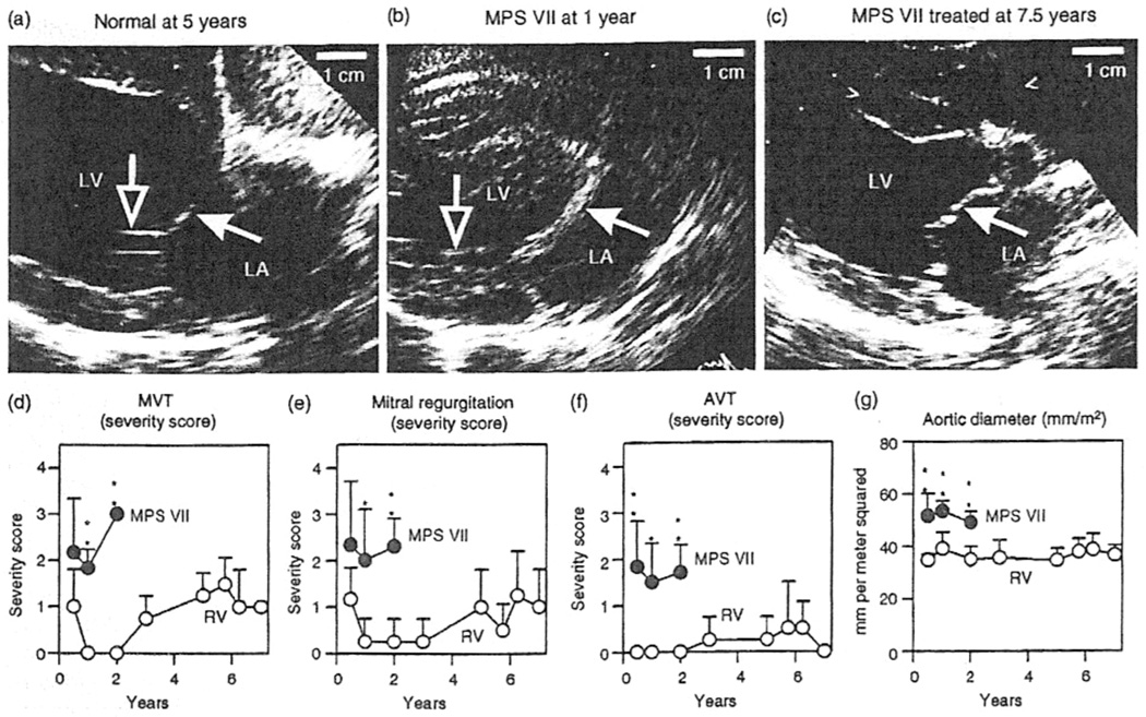
Figure 1.
Echocardiograms in dogs with mucopolysaccharidosis (MPS) VII. Normal dogs, dogs with untreated MPS VII, or dogs with MPS VII treated with retroviral vector underwent echocardiography at the ages indicated above the panels. (a–e) Echocardiographic images of the mitral valve obtained in the long axis from the right parasternum. The cranial (anterior) mitral valve leaflets are indicated by filled white arrows, and the chordae tendinae by open arrows. Note the marked thickening of the valve in the untreated MPS VII dog compared with that in the normal and treated MPS VII dogs. (d–f) Subjective severity score for echocardiogram parameters. Mitral valve thickening (MVT) (d), mitral valve regurgitation (e), and aortic valve thickening (AVT) (f) were scored from 0 (normal) to 4 (severely abnormal) for six or seven dogs with untreated MPS VII and for four dogs with MPS VII treated with retroviral vector, at the indicated ages. Values in the groups at a given age were compared statistically using Student’s t-test (*P = 0.005–0.05; **P = < 0.005). Untreated dogs with MPS VII do not survive beyond 2 years, so it was not possible to compare values in treated dogs with MPS VII with those in age-matched, untreated dogs at older ages. (g) Aortic diameter. The aortic diameter determined in the long axis was normalized to the body surface area (m2), and statistical comparisons were performed as in (d–f). LA, left atrium; LV, left ventricle.

