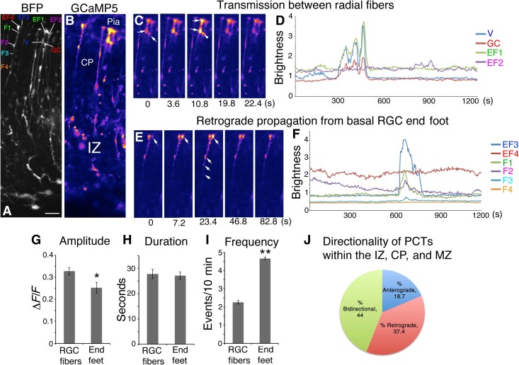
Fig. 2. Calcium transient activity and directionality in RGC fibers and pial end feet.
(A) BFP Z-stack of RGC fibers. (B) Corresponding image of GCaMP5 fluorescence. (C) Time-lapse images of coherent activity in two RGC end feet. (D) Calcium activity traces of the series in (C); note that the multiple fluorescence events of a growth cone (GC) of an adjacent RGC fiber accurately mirror activity of the adjacent end foot. (E) Time-lapse series of pial end foot initiating a Ca2+ transient, which propagates retrogradely through the CP. (F) Calcium activity traces for the series in (E) showing temporal progression of the Ca2+ wavefront in the RGC fiber. (G to I) Population comparison of Ca2+ event properties within RGC fibers and pial end feet showing respective amplitude, duration, and frequency. (J) PCTs originating in the IZ, CP, and MZ categorized by directionality. Note that MZ transients may be nonpropagative or retrograde only. EF1 to EF4, end foot 1 to end foot 4; V, varicosity; F1 to F4, fiber point 1 to fiber point 4. *P < 0.05; **P < 0.001. Error bars, mean ± SEM. Scale bar, 20 μm.

