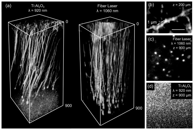
Fig. 5.
(a) Deep imaging comparison between a commercial Ti:S laser tuned to 920 nm and the custom built 1060 nm fiber laser. Image dimensions 400 × 400 × 900 µm3. Fluorescence stacks are pyramidal neurons expressing YFP taken in two separate mice. (b) A zoomed in view of a dendrite at 200 µm depth taken with the fiber laser. Single dendritic spines can be visualized with sub 1 µm features. Scale bar is 8 µm. (c) An image of pyramidal neuron bodies at 900 µm depth taken with the fiber laser. (d) An image at 900 μm depth taken with a commercial Ti:S laser, neuron structures cannot be resolved.

