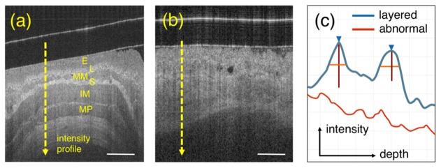
Fig. 4.
Layer detection. (a) shows a representative OCT image of squamous esophagus where the typical layers are visible: squamous epithelium (E), lamina propria (L), muscularis mucosa (MM), submucosa (S), inner muscularis propria (IM) and outer muscularis propria (OM). (b) shows an example of BE tissue where the layers characterizing normal esophagus are absent. (c) schematic A-lines for both layered normal esophagus (from lumen to IM – blue line) and BE (red).. Blue arrow and vertical/horizontal lines correspond to peak position, height and width measured at half-height, respectively. Scale bar equal to 500 µm.

