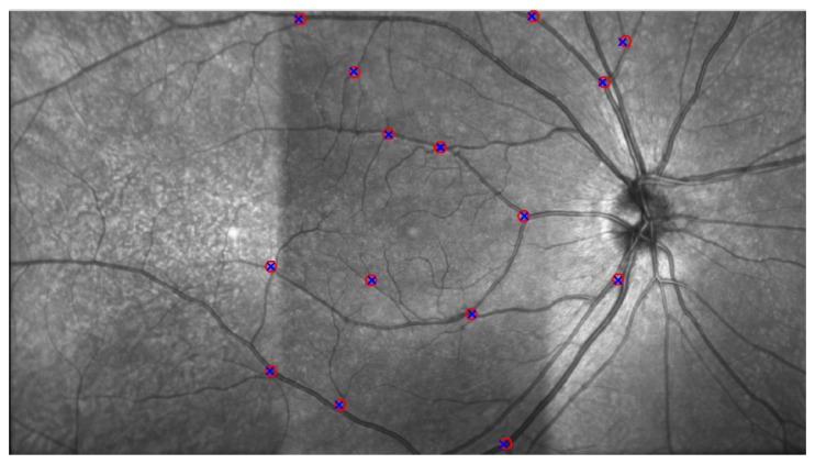
Fig. 3.

A representative example of an aligned and blended SLO image, formed by three overlapping images. A smooth transition of the retinal vasculature can be observed, along with good alignment of the manually selected points used to validate the automatic procedure. Eight landmarks per overlapping region were identified in each of the SLO image-pairs (circle, cross), 16 in total per subject considering both overlapping regions (temporal-central and central-nasal).
