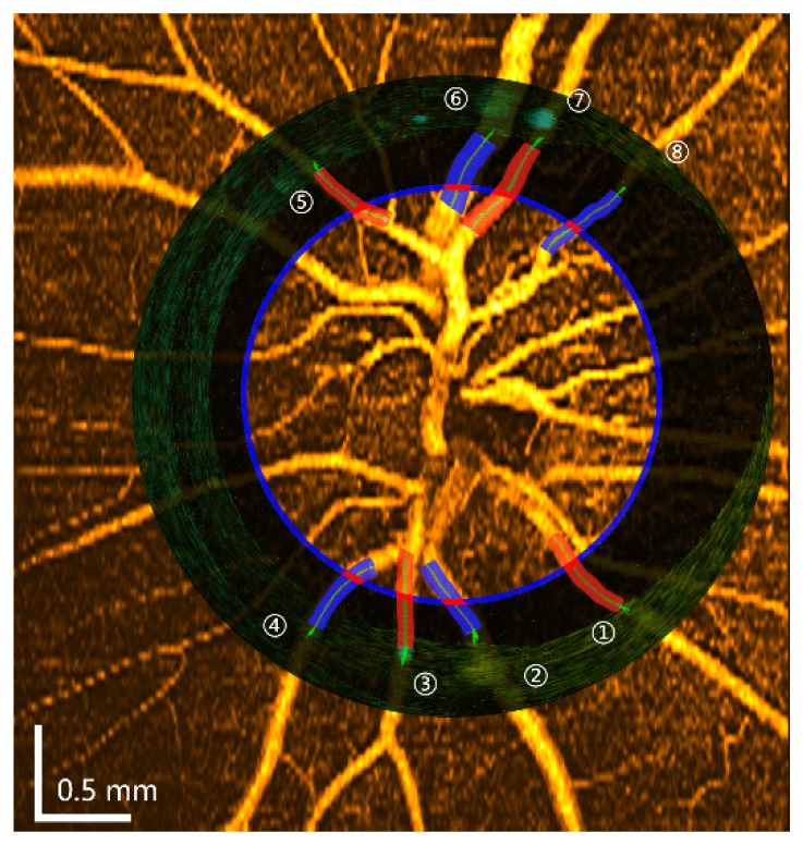
Fig. 5.

Registration between OCT angiography and Doppler OCT. The blue circular line indicates the circular scan position corresponding to the en face view of the OCT angiography image. The circular view of the Doppler image was merged with the angiography image. Eight major vessels were selected, and the locations of the measured vessels were indicated by short red lines. The boundaries of the measured vessels were also overlaid on the angiography image, and the green lines indicate the center of each vessel. The red color indicates arteries and blue color indicates veins.
