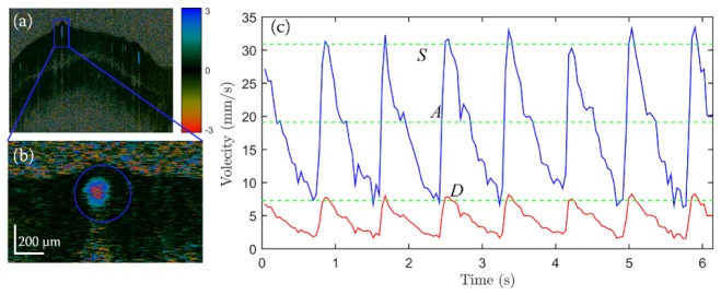
Fig. 8.
Pulsatile flow of a retinal vessel. (a) Doppler OCT image for a circular scan of the optic disc. (b) Enlarged Doppler OCT image of the selected vessel marked by the blue rectangle in (a). (c) Pulsatile velocity profile of the selected vessel within a time span of ~6 s. Red profile, velocity before Doppler angle correction; blue profile, corresponding velocity after correction. Green dashed lines: top, peak systolic velocity (S); middle, average velocity (A), bottom, end diastolic velocity (D).

