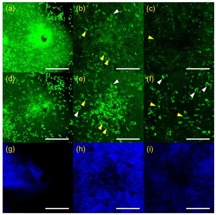
Fig. 5.
3D RCM and TPSHGM images of candida albicans-infected rabbit cornea # 1 in the x-y plane at three depths. (a-c): RCM images at surface, 25, and 60 µm deep from the surface (Visualization 7 (1.3MB, MOV) ), (d-f) and (g-i): corresponding TPSHGM AF and SHG images to the RCM images (Visualization 8 (1.4MB, MOV) , green: autofluorescence, blue: SHG). Scale bar: 100 µm

