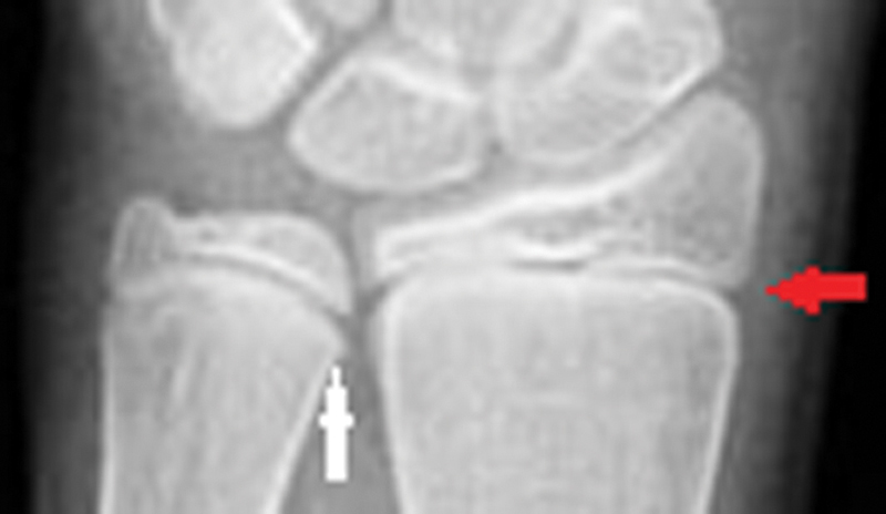
Fig. 1.

Radiograph of the distal radius and ulna showing the R7 grade. Examiners should focus on the medial side capping and the absence of lateral side capping (red arrow). Note the hooklike structure/sharp outgrowth at the medial physis facing the metaphysis that deviates from the physeal line. This radiograph also represents the U6 grade with the appearance of an epiphysis and metaphysis of the same width but no narrowing of the medial physeal plate (white arrow).
