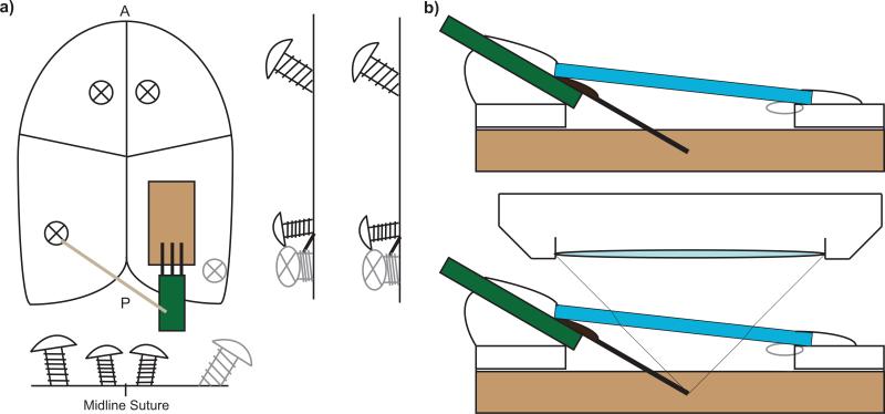
Figure 1.
Scheme for Chronic multiphoton imaging setup. a) Four bone screws are used to anchor the probe and imaging window as well as a well created for a water emersion microscope lens. b) Coverslip windows are gently placed on the probe after filling the cranial space and then sealed. When filling it with aCSF, it is necessary to leave a small air bubble to prevent a leaky seal. Small air bubbles are solubilized in the CSF over the first 12 hours. Probes must be inserted at an angle to prevent collision with the microscope lens. c) Locations of the bone screws. d) Preliminary dental cement well. e) Craniotomy. f) Insertion of the electrode and securing of the reference/ground wire. g) Sealing the craniotomy with Kwik-Sil, coverslip, and dental cement.

