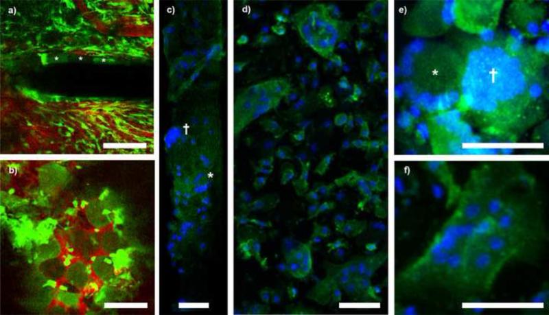
Figure 6.
Foreign body giant cell encapsulation of neural probe and Kwik-Sil at 1 month post implant. A) Large, oval CX3CR1+ cells encapsulated the device with morphology distinct from parenchymal microglia or meningeal macrophages. Cells denoted with asterisks. B) Unidentified round cells also adhered to the Kwik-Sil. C) Explanted electrodes show nuclei patterns (blue, Hoescht stain) consistent with FBGC (†) and Langhans’ giant cells (*). D) Explanted Kwik-Sil with adherent giant cells (Blue: Hoescht; Green: Iba-1). E-F) Identification of FBCG (†)Langhans’ (*), and Touton's giant cells (E) on Kwik-Sil (Blue: Hoescht; Green: Iba1). All scale bars are 50 μm.

