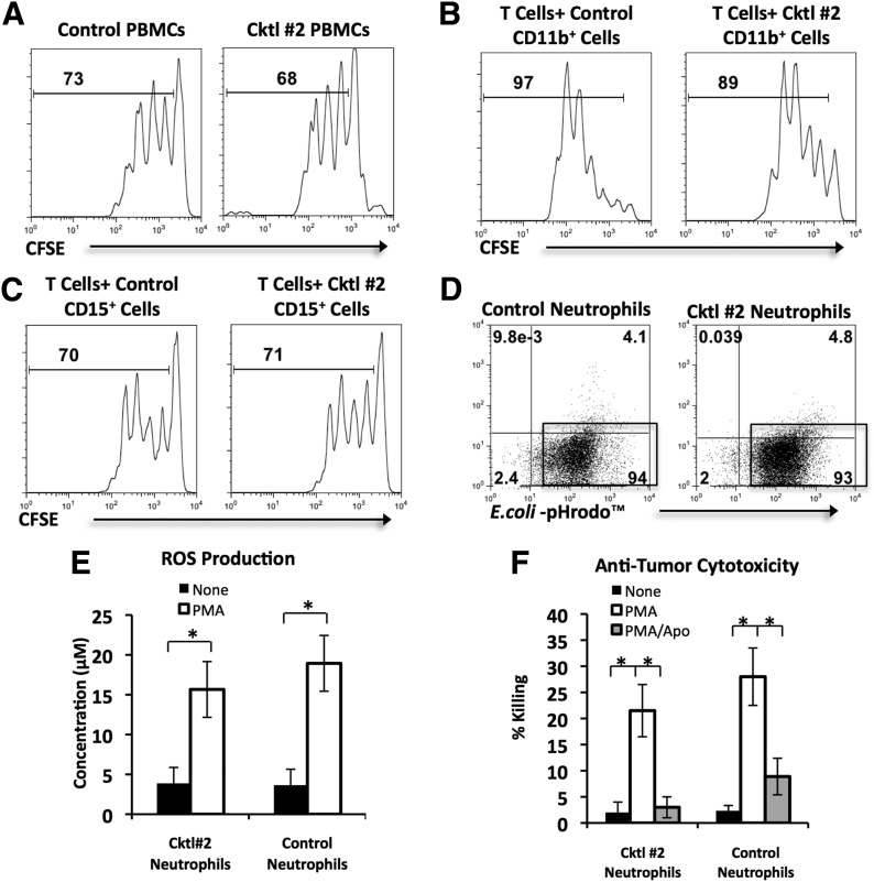
Figure 5. The effect of enzymatic exposure on the functional activity of HD peripheral blood leukocytes.
HD PBMCs and PBNs were incubated with PBS (control) or Cocktail #2 (Coll I, II, IV) for 30 min and then added to multiple in vitro functional assays. For bar graphs, statistical analysis was performed by Student’s t-tests (*P < 0.05). Each bar represents mean ± sem. (A) The effect of enzymatic exposure on T cell proliferation. CFSE-labeled control or Cocktail #2-treated PBMCs were stimulated with plate-bound anti-CD3 antibodies for 4 days. The proliferation of T cells was analyzed by CFSE dilution in the gated CD3+ cells. The histograms, representing 1 experiment of 5, show the percentage of dividing cells. (B and C) The effect of enzymatic exposure on myeloid cell regulation of T cell proliferation. CFSE-labeled T cells were stimulated with plate-bound anti-CD3/CD28 antibodies for 4 days in the presence of control or Cocktail #2-treated CD11b+ or CD15+ cells. The histograms, representing 1 experiment of 5, show the percentage of dividing T cells in the presence of CD11b+ cells or CD15+ cells. (D) Phagocytosis assay. Neutrophils were cocultured for 45 min with E. coli-pHrodo conjugates and then analyzed by flow cytometry. Representative flow cytometry dot plots from 1 of 5 experiments are shown. Percentages of cells that phagocytosed E. coli-pHrodo conjugates are displayed in gates. (E) ROS production by PMA-stimulated neutrophils, which were cultured in the presence or absence of PMA for 1 h before ROS concentration was measured in the supernatant by use of Amplex Red reagent. Graphical summary of 5 experiments is displayed. (F) Anti-tumor cytotoxicity of PMA-stimulated neutrophils. Neutrophils were cocultured in a 1:1 ratio with GFP-expressing A549 human NSCLC cells without PMA, in the presence of PMA, or the presence of PMA/apocynin (Apo) for 16 h. Graphical summary of 5 experiments is displayed.

