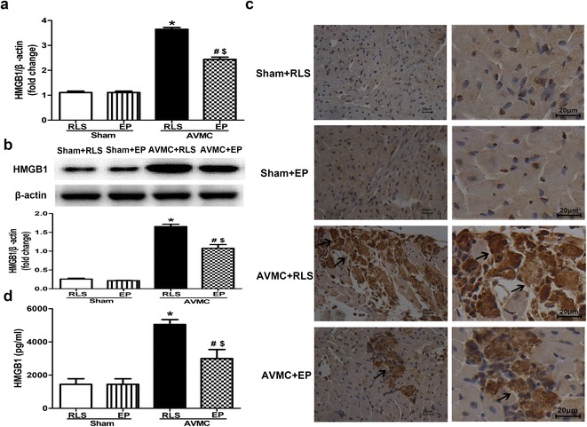
Fig. 4.

Effect of EP on the expression and release of HMGB1. The sham mice and AVMC mice were intraperitoneally administrated with Ringer’s lactate solution (RLS) or 80 mg/kg/day EP from day 5 to day 7 after CVB3 infection. a Real-time PCR analysis of HMGB1 in the hearts. Data were shown as mean ± SE from 6 to 12 individual experiments. *P < 0.001 versus Sham + RLS. #P < 0.001 versus Sham + EP. $P < 0.001 versus AVMC + RLS. b Immunoblot for analysis on HMGB1 expression in the hearts. The blot is showed in the upper part. The quantification of HMGB1 expression was shown in the lower part. Data were shown as mean ± SE from 6 to 12 individual experiments. *P < 0.001 versus Sham + RLS. #P < 0.001 versus Sham + EP. $P < 0.001 versus AVMC + RLS. c Immunochemistry for the localization of HMGB1 in the hearts (Left ×200, ×Right ×400). HMGB1 was predominantly located in the nuclei of the cells in the hearts of sham mice. However, in the hearts of AVMC mice, HMGB1 was not only positive in the nuclei but also positive in the cytoplasm of the cells and extracellular milieu. The black arrows showed abnormal location of HMGB1. d ELISA for quantitative analysis of serum HMGB1. Data were shown as mean ± SE from 6 to 12 individual experiments. *P < 0.001 versus Sham + RLS; # P < 0.05 versus Sham + EP; $ P < 0.01 versus AVMC + RLS
