Figure 3.
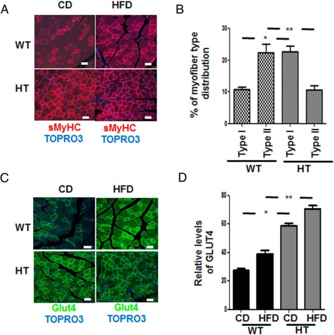
Muscle fiber type switch and enhanced detection of Glut4 in gastrocnemius muscle of HT mice. A, Antimouse sMHC followed by antimouse-labeled 594 confocal immunofluorescence staining of the gastrocnemius muscle of chow-fed (CD) and HFD-fed mice. B, The figure shows the increase in myofiber type switch in the HT mice compared with WT mice. A total of 400 fibers were counted per group (white bar, 40 μm), expression of muscle fibers type I in WT vs HT data represent the mean ± SEM; *, P < .05, and type II WT vs HT; **, P < .002. C, Antirabbit Glut4 followed by antirabbit-labeled 488 in gastrocnemius muscle tissue. Nuclei identification is obtained with TOPRO3; white bar, 40 μm. D, The graph shows the relative levels of GLUT4 receptor in the muscle of HT and WT mice. A total of 300 fibers were counted per group, and a significant expression of GLUT4 was observed in mice fed CD WT vs HT, data represent the mean ± SEM; *, P < .05. A similar significant expression of Glut4 in mice fed HFD between WT and HT; **, P < .001.

