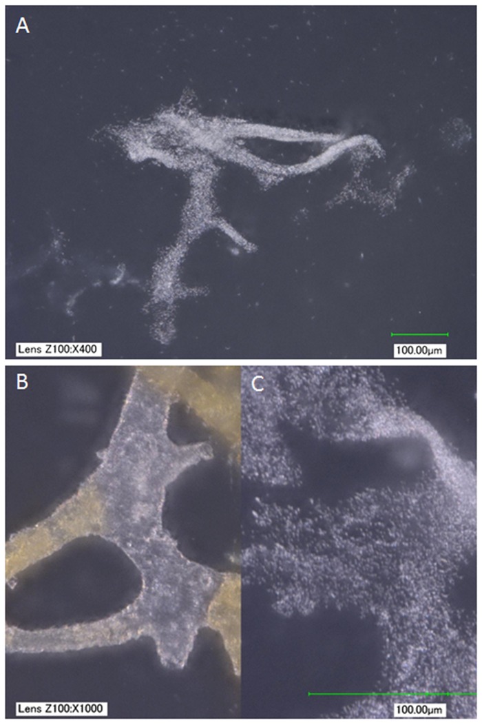
Fig 3.
(A) B.cereus biofilm grown in cow bone from which organics had been removed (see methods). (B, C) Side-by-side comparison of vessels from MOR 2967-C5-1, a second Brachylophosaurus specimen from similar deposits (B), and (C) B. cereus biofilm recovered from cow bone at the same magnification. At low magnification, the biofilm mimics vessel shapes; higher magnification reveals that the biofilm is not hollow, as are the vessels, but rather amorphous clusters of cells. In addition, a red substance is clearly visible and differentially distributed within the hollow dinosaur vessels; no similar features are seen in the biofilm. Images taken with a KEYENCE VHX-2000 digital microscope, scale bar as indicated.

