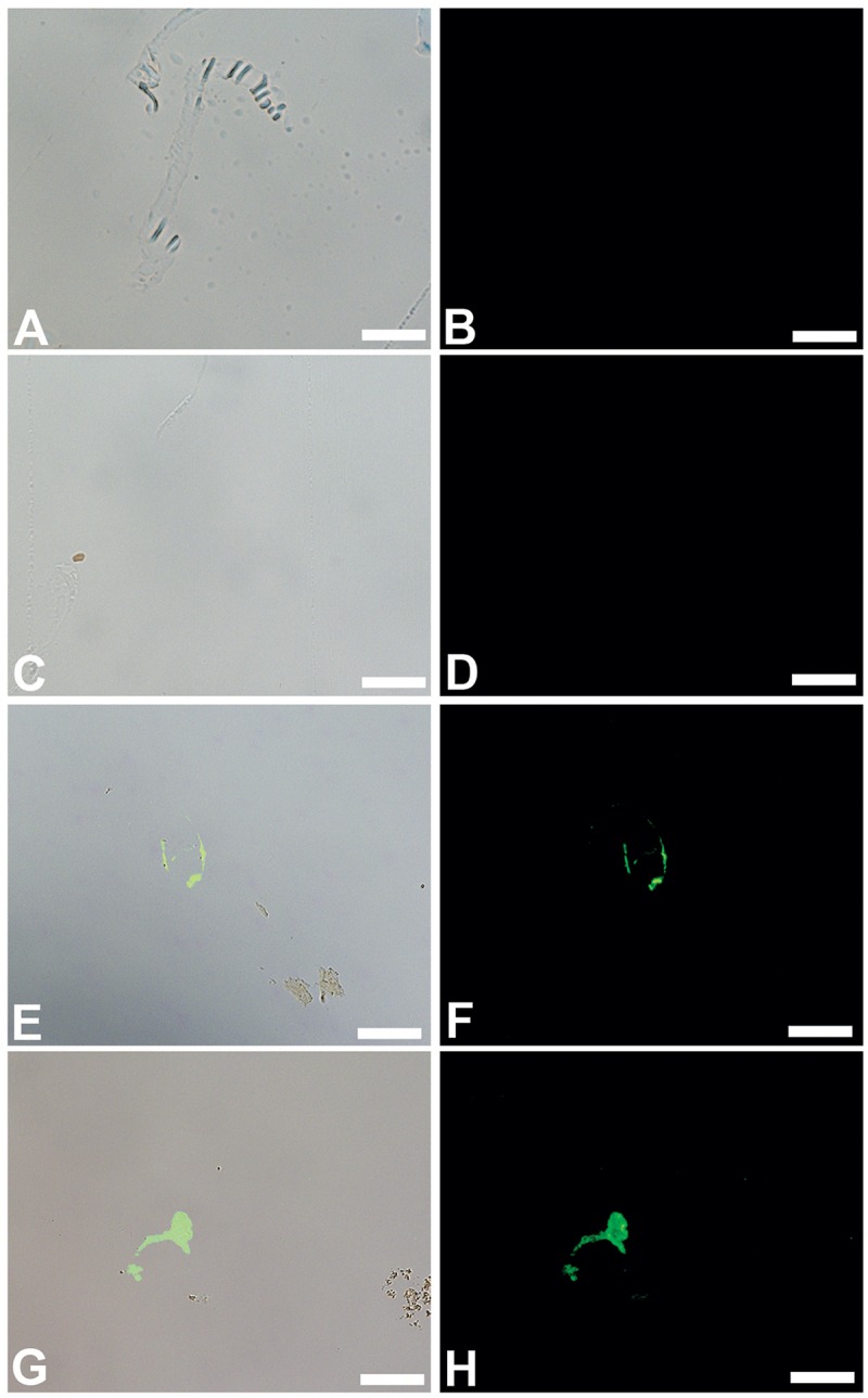
Fig 8.

Overlay and fluorescent images of B. cereus (A, B), S. epidermidis (C, D) B. canadensis vessels (E, F) and T. rex vessels (G, H) exposed to antiserum raised against purified ostrich hemoglobin. No binding of these antibodies to either biofilm is visualized, but specific binding to vessels from both dinosaurs is seen. Scale bar for all images = 20 μm
