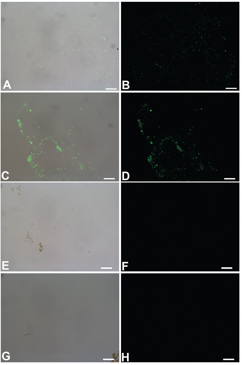
Fig 9.

Overlay and fluorescent images of B cereus (A, B), S. epidermidis (C, D) biofilms, B. canadensis (E, F) and T. rex (G, H) vessels, exposed to antibodies raised against peptidoglycan, a bacterially produced glycosaminoglycan that is a component of both bacterial cell walls and the EPS they secrete [33]. Binding of these antibodies is evident in both biofilm products, but no binding is seen of these antibodies to either dinosaur vessel. Scale bar for all images = 20 μm
