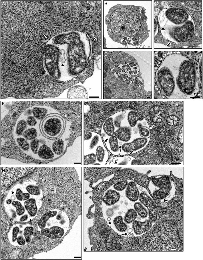
Figure 9.
The ApV interacts with the ER as visualized by TEM. A. phagocytophilum infected HL-60 cells were examined by TEM. White hatched boxes denote areas in the images presented in (B,D) that are presented as enlarged panels in (C,E), respectively. The white arrow in (A) denotes a RER-ApV contact site. Black arrows in (A,C,G) denote autophagosomes contacting the ApV membrane. Black arrowheads in (A,C,G), and (I) demarcate autophagic bodies present within the ApV lumen. White arrowheads in (E) denote ribosomes that label the cytosolic face of the ApV membrane. Thin white arrows in (F,G) demarcate membranes within the ApV lumen associating with A. phagocytophilum organisms. Thin black arrows in (G,H,I) point to vesicles within the ApV lumen in close apposition to A. phagocytophilum bacteria. Scale bars, 0.5 μm. Results shown are representative of two experiments in which a combined total of over 200 different electron micrographs were analyzed.

