
Figure 1. PC X‐ray and angiography images .
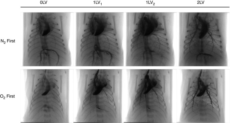
Representative X‐ray image sequences of newborn rabbits imaged prior to ventilation (0LV), following unilateral ventilation of the right lung (1LV1) with either 100% N2 or 100% O2, subsequent ventilation with air (21% O2) in all kits (1LV2), and later ventilation of both lungs in air (2LV). Images were obtained 1–3 s following iodine bolus injection.