
Figure 4. Blood vessel internal diameter changes during ventilation .
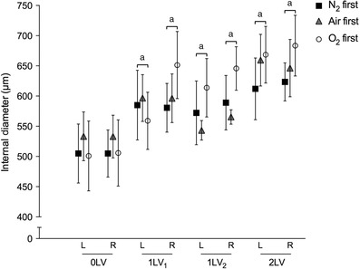
Mean internal diameter (μm,± SEM) of the left and right axial arteries at the seventh intercostal space at each ventilation period (0LV, 1LV1, 1LV2 and 2LV) in the N2 first (black solid squares), air first (grey solid triangles) and O2 first (open circles) groups. a P < 0.05 compared to baseline (0LV) in the same lung in the same group.