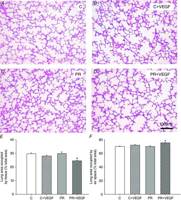
Figure 6. Evaluation of total percentage area occupied by tissue and air space in the fetal lung .

Micrographs of hematoxylin and eosin staining in the lung tissue of the Control (C) (A), C + VEGF (B), PR (C) and PR + VEGF (D) fetuses (100× magnification, scale bar = 100 μm). Percentage of total area occupied by tissue (E) and air space (F) in the fetal lung determined by point counting in the C (n = 9, open bar), C + VEGF (n = 7, open hashed bar), PR (n = 7, grey bar) and PR + VEGF (n = 3, grey hashed bar) fetuses. VEGF administration significantly decreased the total percentage of tissue and increased the total percentage of air space in the PR lung, but not the Control lung (P = 0.06). Data (mean ± SEM) were analysed by two‐way ANOVA for treatment (Control vs. PR) and drug (Saline vs. VEGF). *P < 0.05 from Control or PR (i.e. drug effect).
