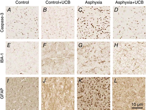
Figure 3. Representative photomicrographs of the thalamic nuclei stained for apoptosis, inflammation and astrogliosis .

Apoptosis: control (A), control+UCB (B), asphyxia (C) and asphyxia+UCB (D); inflammation: control (E), control+UCB (F), asphyxia (G) and asphyxia+UCB (H); and astrogliosis: control (I), control+UCB (J), asphyxia (K) and asphyxia+UCB (L). Asphyxia resulted in increased apoptosis (activated caspase‐3), inflammation (IBA‐1) and astrogliosis (GFAP) compared to controls. UCB administration to asphyxia animals resulted in significantly reduced numbers of apoptotic cells, inflammatory cells and astrocytes. UCB administration to control animals did not result in any change from control only.
