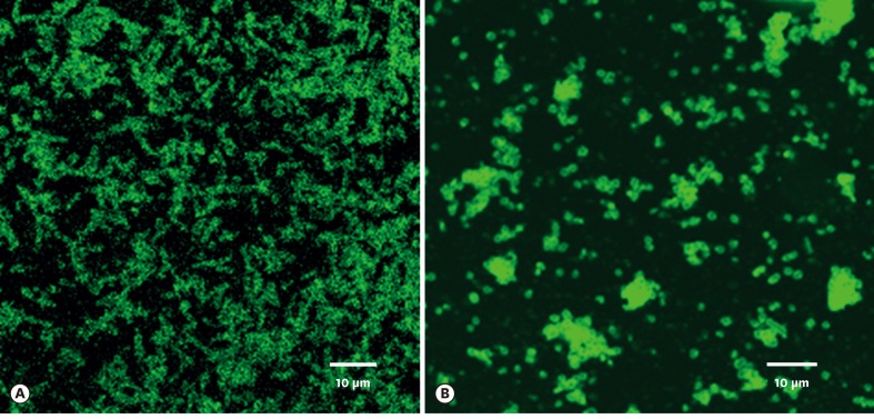
Figure 1.
Surface proteins of P. gingivalis and T. forsythia bound to T. forsythia (A) and P. gingivalis cells (B), respectively, as observed under epifluorescence microscopy using FITC-conjugated streptavidin antibody. Fluorescence-labeled rod-shaped T. forsythia cells are clearly evident (A). Fluorescence-labeled coccobacilli-shaped P. gingivalis cells can be seen (B). For details, refer to the main text.

