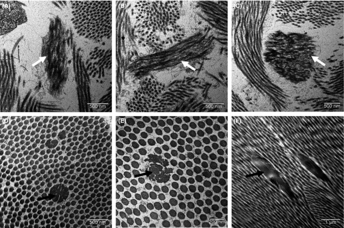
Figure 2.

Electron microscopy analysis of skin biopsy: fragmented and frayed elastic fibers (A, B, and C, white arrows), abnormal collagen fibers (black arrows) with a wide variability of fiber caliber and the presence of cauliflower‐like fibers (D, E, F).
