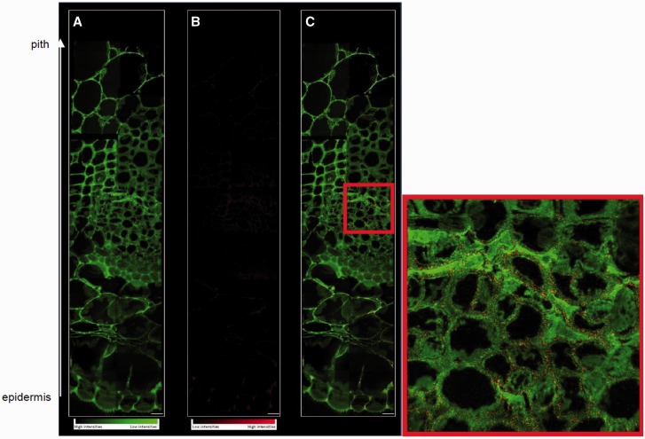
Fig. 9.
NanoSIMS analysis of the peripheral region to the central region of a basal stem section prepared by freezing followed by freeze substitution. (A) 12C14N– image in green scale color from low to high intensity, (B) copper distribution 63Cu– in red scale color, (C) color overlay for CN and Cu. Scale bar = 5 µm. In the red square is a focus of the cambial region of the stem.

