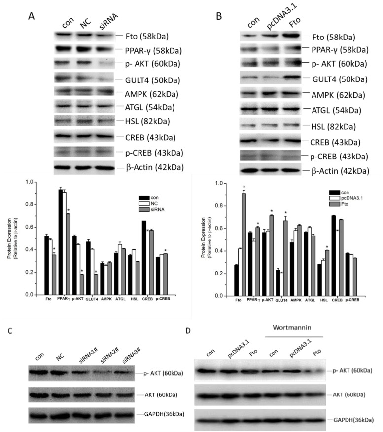
Figure 5.
Effect of Fto on proliferation and differentiation of pre-adipocyte. (A) 3T3-L1 cells transfected with/without NC siRNA or Fto siRNA, as well as (B) Fto recombinant plasmid or vector “mock” transfected 3T3-L1 cells were harvested 24 h after transfection for protein extraction and quantified. Fifty microgram of total protein were analyzed by Western blot. Expressions and activities of specific transcription factors and signaling factors were detected. The expression level was displayed as fold changes in band density. Results are expressed as the means ± SD from three independent experiments. * p < 0.05, vs. the mock transfection group. (C) Further, three different Fto siRNA were transfected into 3T3-L1 cells for 24 h. The phosphor-Akt and Akt expression were examined by Western blot assay. (D) 3T3-L1 cells were transfected with vector plasmid and Fto plasmid for 24 h, then were cultured in the absence or presence of the PI3k inhibitor (Wortmannin, 100 nmol/L) for 24 h. The expression level of phospho-Akt and Akt was determined by Western blot analysis.

