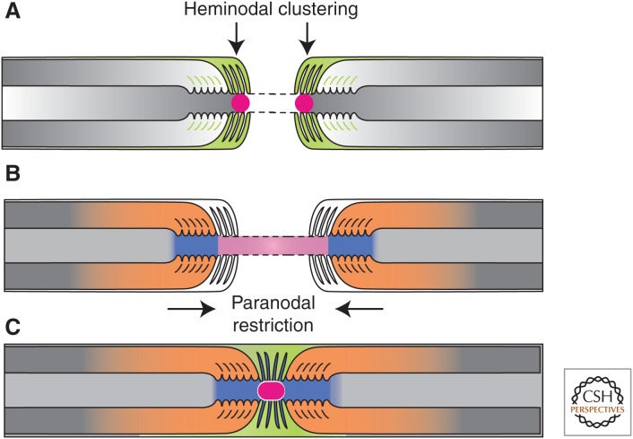
Figure 3.
The formation of PNS nodes of Ranvier. (A) Na+ channels (red circle) are trapped at heminodes that are contacted by Schwann cell microvilli ([MV] orange). Axon–glia interaction at this site is mediated by binding of gliomedin and glial NrCAM to axonal NF186. A transmembrane form of glial NrCAM traps gliomedin on Schwann cell microvilli and enhances its binding to axonal NF186. (B) The distribution of Na+ channels is restricted between two forming myelin segments by the PNJ (blue). Three CAMs, NF155 present at the glial paranodal loops, and an axonal complex of Caspr and contactin mediate axon–glia interaction and the formation of the PNJ. (C) These two extrinsic cooperating mechanisms provide reciprocal backup systems and ensure that Na+ channels are found at high density at the nodes. Finally, the entire nodal complex requires stabilization and interaction with cytoskeletal and scaffolding proteins, which constitute a third, intrinsic mechanism for Na+ channel clustering. (From Feinberg et al. 2010; reprinted, with permission, from Elsevier Limited © 2010.)

