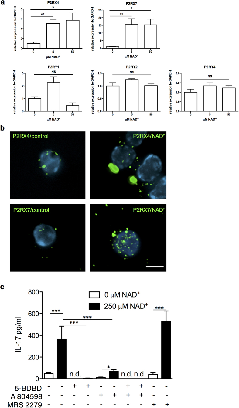
Figure 4. NAD+ signals through P2RX4 and P2RX7 receptors.
CD4+ CD25+ Tregs were cultured in the presence of α-CD3, α-CD28, and IL-2 with increasing concentrations of NAD+; after 24 hrs (a) mRNA levels for P2RX4, P2RX7, P2RY1, P2RY2, and P2RY4 were measured by real-time PCR (n = 6 per group). (b) Freshly isolated T cells were cultured for 24 hrs in presence of vehicle alone (right column) or 50 μM NAD+ (left column). Cells were collected and stained at 4 °C for either P2RX4 (top row) or P2RX7 (bottom row) without prior fixation or permeabilization. Stacks of 20, 2-channel images were acquired under each condition; resulting stacks were de-convolved and re-constituted for further analysis. Results show that treatment of T cells with NAD+ increased the cell surface expression levels of both receptors and in the case of P2RX4, NAD+ promotes capping-like distribution pattern. (c) Tregs were cultured as described above with or without NAD+ (250μM) and with or without MRS 2279 (a selective antagonist of P2RY1), 5-BDBD (a selective antagonist of P2RX4) or A 804598 (a selective antagonist of P2RX7) and IL-17A secretion was measured by ELISA (n = 6 per group). Data derived from two independent experiments. Data represent mean ± s.d. *P < 0.05; **P < 0.01; ***P < 0.001. ANOVA tests were used to compare groups.

