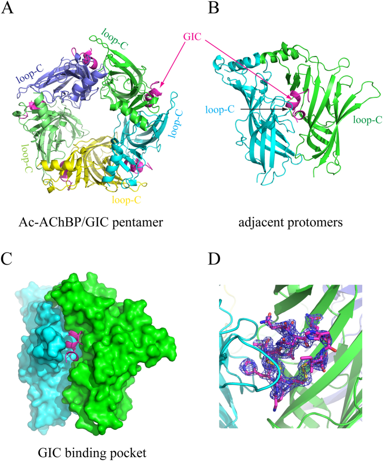
Figure 1. The X-ray crystal structure of Ac-AChBP in complex with α-conotoxin GIC.
(A) The top view of the pentameric structure with five Ac-AChBP protomers, each in different colours and five α-conotoxin GIC molecules in magenta. (B) The side view of two adjacent protomers of the pentamer with a bound α-conotoxin GIC molecule (in magenta). (C) The side view of the surface model of two adjacent protomers with a bound α-conotoxin GIC molecule (in magenta) inside the binding pocket. (D) Fo-Fc electron density omit map contoured at 3.0 σ surrounding the GIC.

