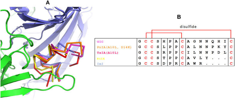
Figure 2. Comparison of different α-conotoxins bound by Ac-AChBP.
(A) Backbone orientations observed in co-crystal structures of Ac-AChBP with five α-conotoxins. The backbone of GIC is shown in magenta, PnIA (A10L, D14K) in orange, ImI in light blue, TxIA (A10L) in red and BuIA in yellow. (B) Multiple sequence alignment of α-conotoxins GIC, PnIA (A10L, D14K), TxIA (A10L), ImI and BuIA. Disulfide bridges between Cys2-Cys8 and Cys3-Cys16 are shown in red.

