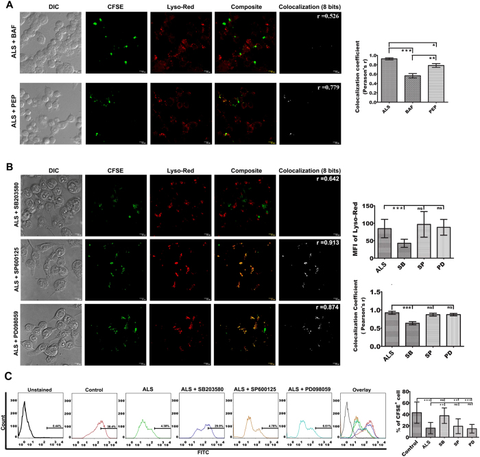
Figure 7. Involvement of p38 MAPK in phagosome maturation.
(A) MΦs were infected with ALS for 4 h, washed, and incubated for further 8 h in fresh media. These MΦs were then infected with CFSE labeled PLD for 4 h and then treated with bafilomycin A1 and pepstatin A for 4 h and then washed, fresh media was added. After 12 h post infection, co-localization of LysoRed and CFSE labeled PLD was observed under a microscope (left panel). Co-localization index (Pearsons’ r) were graphically represented (right panel). (B) RAW 264.7 cells were infected with ALS for 4 h, washed, and fresh media was added and incubated for additional 12 h. These MΦs were then infected with CFSE labeled PLD for 4 h and then treated with SB203580, SP600125, and PD098059 for another 4 h. Cells were then washed and incubated for further 8 h in fresh media. Co-localization of LysoRed and CFSE labeled PLD was observed under microscope (left panel). Co-localization index (Pearsons’ r) were graphically represented (right panel).(C) MΦs were primed with ALS, infected with PLD and treated with MAPK inhibitors as stated above. Viable parasite load was determined by flowcytometry (left panel). Percent positive cells were represented graphically (right panel). Data are representative as the mean ± SD and are the cumulative results of three independent experiments *p < 0.05, **p < 0.01, ***p < 0.001.

