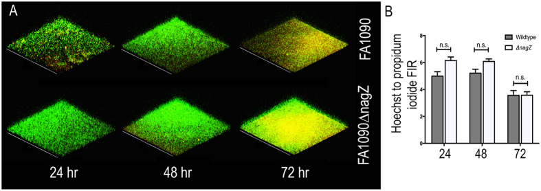
Figure 4. Viability of cells in gonococcal biofilms.
(A) Static biofilms were formed with FA1090 and FA1090∆nagZ strains for different periods of time, and visualized by confocal microscope after staining with propidium iodide (red) and Hoechst (green). (B) The fluorescence intensity ratio (FIR) between Hoechst and PI was measured for both strains at the three time points. No significant differences were observed between the ration of dead and live cells at the various time points.

