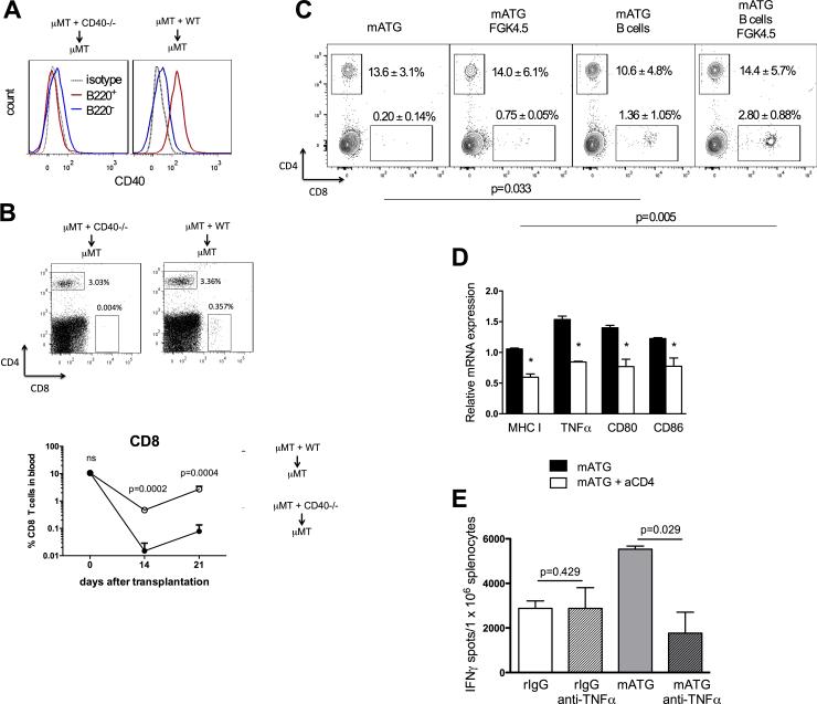
Figure 7. CD40 expression on B cells and TNFα are required for CD8 T cell reconstitution.
A-B. μMT + CD40−/− ➔ μMT (lack CD40 only on B cells) and μMT + WT ➔ μMT (express CD40) mixed bone marrow chimeras were generated as described in the Methods section. CD40 expression on B220+ vs B220− cells was evaluated by flow cytometry at four weeks after bone marrow reconstitution (A). The chimeric mice were transplanted with BALB/c heart allografts and treated with mATG on days 0 and 4 after transplantation. The percentages of CD4+ and CD8+ T cells in peripheral blood were determined by flow cytometry on days 14 and 21 after transplantation (B). Representative histograms (A), dot plots (B, top) and mean ±SD (B, bottom) are shown for 3-4 animals/group. C. B6.CD40−/− mice were transplanted with BALB/c heart allografts and treated with mATG on days 0 and 4 posttransplant. Splenic B cells from naïve B6 mice were adoptively transferred into recipients on day 5 posttransplant. Groups of recipients with or without B cell transfer were injected with agonistic anti-CD40 mAb FGK4.5 on days 6 and 7. The percentages of CD4 and CD8 T cells in blood were analyzed on day 42 by flow cytometry. P values are shown to compare percentages of CD8+ T cells. D. Non-transplanted B6 mice were treated with mATG (d. 0 and 4) with or without anti-CD4 mAb. Spleen B cells were isolated on day 14 after first mATG injection and mRNA expression was analyzed by qRT-PCR. E. B6 recipients of BALB/c heart allografts were treated with mATG or rIgG alone or in combination with anti-TNFα Ab (XT3.11, 0.5 mg i.v. on d. −1 and d. 1). Anti-donor IFNγ production by recipient spleen cells was measured at d. 10 posttransplant. All panels represent mean ±SD for 4-9 mice/group analyzed at each time point. *P < 0.05; **P < 0.01; ***P < 0.001.

