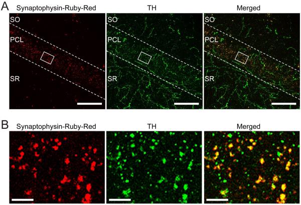
Figure 2. Dopaminergic terminals and axons in the CA1 show high co-localization with tyrosine hydroxylase (TH).
(A) Confocal images of the CA1 field of a Slc6a3ires-cre/+ mouse injected with Synaptophysin-Ruby-Red virus in the VTA/SNc area. Images show punctate Synaptophysin-Ruby-Red signal (in red) mainly along the pyramidal cell layer of CA1 and TH immunoreactivity (in green). A merged overlay of the two signals, shows a high degree of co-localization (yellow-orange) of Synaptophysin-Ruby-Red with TH (Ruby-Red/TH double-positive: 93 ± 2%, n = 6) providing further evidence of the existence of midbrain dopaminergic innervation of the hippocampus. SO: stratum oriens, PCL: pyramidal cell layer, SR: stratum radiatum. Scale bars, 50 μm. (B) High magnification images of the white boxed area in (A), showing individual and composite images of the different labels including the co-localization (Merged). Scale bars, 5 μm.

