Figure 2.
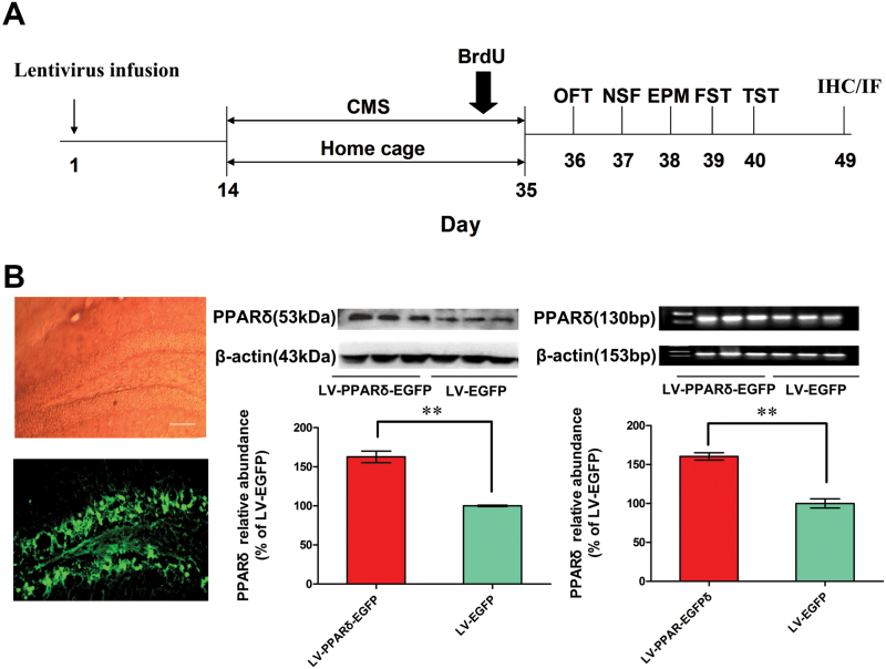
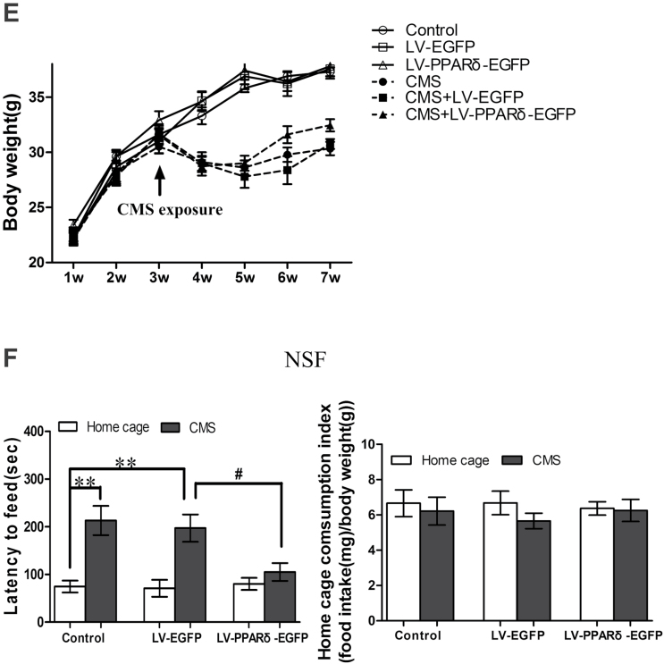
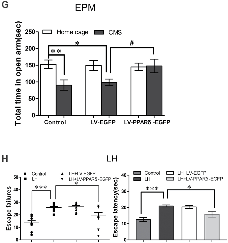
Hippocampal peroxisome proliferator-activated receptor δ (PPARδ) overexpression decreases depressive behaviors. (A) Schematic timeline of the experimental procedure. OFT, open field test; NSF, novelty-suppressed feeding; EPM, elevated plus maze; FST, forced swimming test; TST, tail suspension test; IHC, immunohistochemistry; IF, immunofluorescence. (B) Shown are representative dentate gyrus (DG) area with transfection of a lentiviral vector that selectively expresses PPARδ with enhanced green fluorescent protein (LV-PPARδ-EGFP), PPARδ protein, or mRNA in the hippocampus (n = 3). Shown are the (C) immobility time in the forced swim test (FST) and tail suspension test (TST), (D) physical state index, (E) body weight, (F) latency to feed and home cage consumption index in NSF test, and (G) total time spent in open arms in EPM in the mice intrahippocampally microinjected with LV-PPARδ-EGFP (2×10⁹ TU, 2μl/side), followed 2 weeks later by chronic mild stress (CMS) for 21 d (n = 8–10). (H) Shown are escape failures and escape latency in the learned helplessness (LH) test (n = 8–10). Data are mean±standard error of the mean. (B) ** p < 0.01 compared with a lentiviral vector expressing EGFP alone (LV-EGFP); (C-G)* p < 0.05, ** p < 0.01 compared with control; # p < 0.05, ## p < 0.01, compared with CMS plus LV-EGFP; (H) * p < 0.05, ** p < 0.01 compared with LH.


