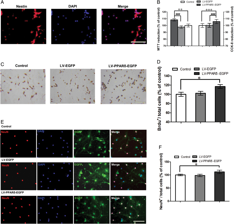
Figure 4.
Peroxisome proliferator-activated receptor δ (PPARδ) overexpression promoted proliferation and differentiation of neural stem cells (NSCs) in vitro. (A) Shown is the protein marker nestin, expressed by NSCs. (B) The coding sequence of mouse PPARδ (LV-PPARδ) increases proliferation determined by 3-(4, 5-dimethythiazole-2-yl)-2, 5-diphenyl-tetrazolium bromide (MTT) and cell counting kit (CCK-8) assays (n = 6). (C) Representatives of bromodeoxyuridine positive (BrdU+)-labeled cells of the NSCs after treatment with a lentiviral vector that selectively expresses PPARδ with enhanced green fluorescent protein (LV-PPARδ-EGFP). (D) Statistical graph shows the number of BrdU+ cells in different groups (n = 6). (E) Immunocytochemistry for neuronal marker neuronal nuclear antigen (NeuN) was used to assess neural differentiation in NSCs treated with LV-PPARδ. (F) Shown are statistical data of NeuN+-positive neurons in different groups (n = 6). Data are mean ± standard error of the mean. * p < 0.05, ** p < 0.01, *** p < 0.001, compared with control; # p < 0.001, compared with a lentiviral vector expressing EGFP alone (LV-EGFP). Scale bars = 50 μm.

