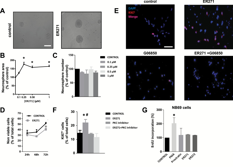
Figure 6.
Effect of ER271 (2) on neural progenitor cell (NPC) proliferation. (A) Phase contrast microscopy images of neurosphere cultures stimulated with basic fibroblast growth factor (bFGF) in the absence and presence of 1 µM ER271 (2). Calibration bar indicates 200 µm. (B) Effect of different concentrations of ER271 (2) on neurosphere area. (C) Effect of different concentrations of ER271 (2) on neurosphere number. (D) Effect of 1 µM ER271 (2) on cell viability. (E) Fluorescence microscopy images of NPC cultures stimulated with bFGF in the presence and absence of 1 µM ER271 (2) and/or 0.5 µM of the protein kinase C (PKC) inhibitor G06850. Cells were immunostained for Ki67 detection and nuclei were counterstained with 4',6-diamidino-2-phenylindole. (F) Quantification of Ki67+ cells in cultures described in E. (G) Bromodeoxyuridine (BrdU) incorporation into NB69 human neuroblastoma cells (see supplementary file for procedure details) cultured for 48 hours in the presence of 8nM phorbol-12-myristate-13-acetate (PMA), 5 µM prostratin, 1 µM ER271 (2), 1 µM ER272 (1), or none (control). #P<.05 compared with the other groups by 1-way ANOVA.

