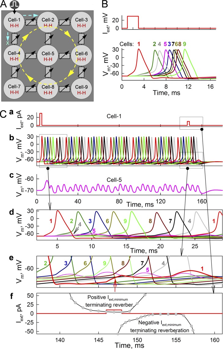
Figure 7.
Formation of reverberation of excitation in a cluster of nine neurons connected through GJs modulatable by an MC16SM. (B) Spread of APs from stimulated cell 1 through all cells shown in A. gj values of GJs varied randomly from 0.175 to 0.2 nS and did not exhibit I-V rectification. (C) Initiation and termination of reverberation of excitation (b) by a single stimulus applied to cell 1 at 2 and 146 ms, respectively (a). Cell 5 is located at the core of reverberation and exhibits only electrotonic depolarizations not transforming into APs (c). The first stimulus caused AP, which spreads to cell 2 but not to cell 4 (d). Reverberation of excitation was stopped by the second pulse, which caused depolarization of ∼3 mV in the middle of cell 1 cycle (red arrow in e) and prevented reexcitation of cell 1 by cell 4. (f) Summarized data demonstrating time windows during which positive and negative Iext,cell 1 pulses of minimal amplitude were able to terminate reverberation of excitation.

