Figure 3.
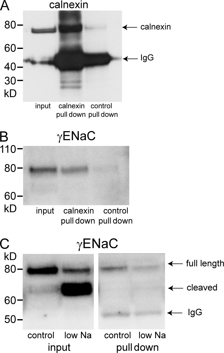
Pull-down of membrane proteins using anti-calnexin antibodies. Calnexin-containing microsomes from rat kidneys were partially purified using differential and density-gradient centrifugation (see Materials and methods). These fractions were incubated with protein G–coated beads primed with anti-calnexin antibody. (A) Significant amounts of calnexin were pulled down from the starting material (input) when beads were coated with anti-calnexin but not with control antibody. The bands at 55 kD represent the IgG species used for pull-downs. (B) Significant amounts of γENaC were pulled down from the starting material with anti-calnexin, but not with control antibody. Only the full-length species was detected. (C) Pull-down of membranes from kidneys of control and Na-depleted rats. The starting material contains both full-length and cleaved γENaC, with the full-length predominating under control conditions and the cleaved predominating after Na depletion. The membranes isolated with anti-calnexin contain only full-length subunits. The abundance decreased with Na depletion. The 55-kD bands represent the anti-calnexin IgG used for the pull-down. The results are representative of four independent experiments. Quantification is shown in Fig. 7.

