Figure 7.
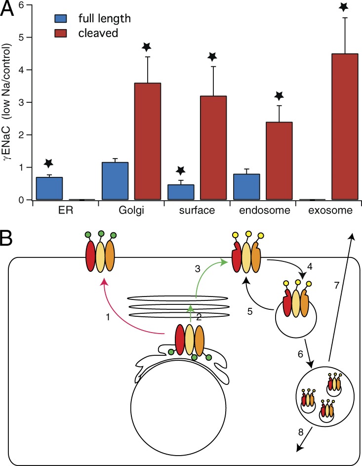
Fractional changes in full-length and cleaved γENaC in various membrane compartments in kidneys from Na-depleted versus control rats. (A) Compartments were identified by calnexin pull-down (ER, n = 4), GM130 pull-down (Golgi, n = 3), surface biotinylation (plasma membrane, n = 4), Rab11 pull-down (endosomes, n = 4), and exosomes (n = 8). Data are represented as mean ± SEM. *, P < 0.05 by t test. (B) Cartoon showing possible trafficking steps stimulated (green) or inhibited (red) by aldosterone/Na depletion. Step 1 is a putative alternative route from ER to the apical membrane that bypasses the final glycosylation and furin-dependent proteolysis of the Golgi apparatus. It appears to be slowed with Na depletion. Step 2 is transfer from ER to Golgi and may be stimulated by Na depletion. Step 3 is trafficking of cleaved and glycosylated protein from Golgi to apical membrane; it is accelerated by low Na. Steps 4 and 5 represent retrieval of protein into endosomes and recycling back to the surface. We cannot evaluate the effects of low Na on these steps. Endosomal retrieval is expected to increase caused by increased density of channels in the apical membrane. Steps 6 and 7 represent transfer to multivesicular bodies and release into the urine as exosomes. These rates also may be increased by the amount of protein in the endosomes. Step 8 represents eventual degradation.

