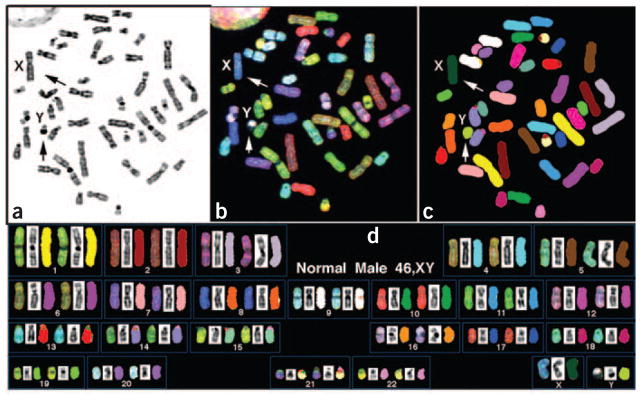
Figure 4.
Normal human male (46 chromosomes, XY) metaphase spread analyzed by SKY. (a) Inverted-DAPI image of a chromosome metaphase spread. The banding pattern is similar to G-banding but accentuates the heterochromatic region of some chromosomes (for example, 1, 9, 16 and Y). (b) Same metaphase with RGB display colors. (c) Same metaphase spread with classification pseudo-colors. (d) Karyotype of the same metaphase spread. Arrows in panels a–c identify the X and Y chromosomes.

