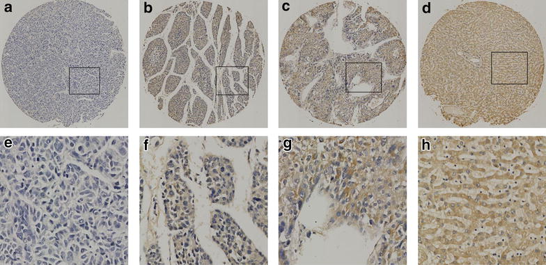
Fig. 2.

The expression of SUSD2 protein in HCC tissues and normal liver tissues by IHC on the TMA. a Weak staining of SUSD2 was detected in a HCC tissue. b Moderate staining of SUSD2 was detected in a HCC tissue, in which more than 70 % of HCC cells stain positively for SUSD2 protein in the cytoplasm. c Strong staining of SUSD2 was detected in a HCC case, in which more than 90 % HCC cells showed positive staining of SUSD2 protein in the cytoplasm. d Strong staining of SUSD2 was detected in a normal liver tissue, in which about 100 % cells showed positive staining of SUSD2 protein in the cytoplasm. e–h demonstrate the higher magnification (400×) from the area of black square in (a–d), respectively
