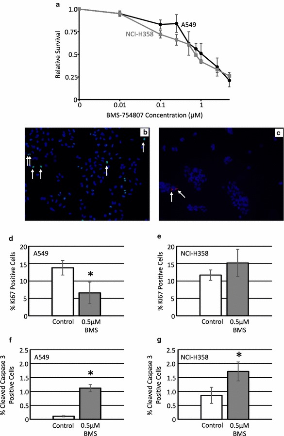
Fig. 2.

Treatment with BMS-754807 reduces cell viability in a concentration dependent manner. A549 (black line) and NCI-H358 (grey line) cells were treated with various concentrations of BMS-754807 every 24 h for 72 h. Cell viability was then measured with a WST-1 assay (a; n = 3). Immunofluorescence for Ki67 and cleaved caspase 3 were used to evaluate the impact of BMS-754807 on these cellular properties. A representative image of Ki67 (b; green color) and cleaved caspase 3 (c; red color) are presented as overlay images of cell nuclei stained with 4′,6-diamidino-2-phenylindole (DAPI, blue color). Arrows highlight some of the positive cells in each image. The number of Ki67 positive cells (d, e) and cleaved caspase 3 positive cells (f, g) along with the total number of cells were counted 24 h after treatment with 0.5 μM BMS-754807 and are presented as relative proliferation (d, e) or relative apoptosis (f, g) in A549 (d, f) and NCI-H358 (e, g) cells. The data is presented as mean ± SEM (n = 4) and the percentage of positive cells have been normalized to the DMSO control. *p < 0.05 as determined by a paired Student’s T-test
