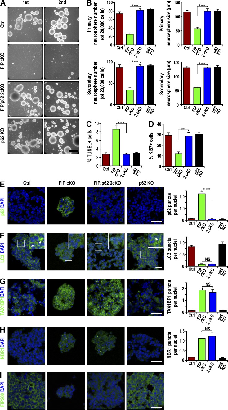
Figure 5.
Defective self-renewal of Fip200-deficient NSCs in vitro restored by p62 KO. (A) Primary (first) and secondary (second) neurosphere formation from Ctrl, Fip200GFAP cKO, 2cKO, and p62 KO mice at P28. Representative phase contrast images. (B) Mean ± SEM of the number and size of primary and secondary neurospheres (n = 3 mice each). (C and D) Mean ± SEM of the percentage of TUNEL+ (C) and Ki67+ (D) cells in neurospheres from Ctrl, Fip200GFAP cKO, 2cKO, and p62 KO mice at P28 by immunofluorescence (n = 3 mice each, >500 cells per mouse). (E–H) Immunofluorescence of p62 (E), LC3 (F), TAX1BP1 (G), NBR1 (H), Fip200 (I), and DAPI in neurospheres from Ctrl, Fip200GFAP cKO, 2cKO, and p62 KO mice at P28. Mean ± SEM of p62 aggregates (E), LC3 puncta (F), TAX1BP1 aggregates (G), and NBR1 aggregates (H) per neurosphere cell. Boxed areas in F show more detail for LC3 puncta, marked by arrows in insets (n = 3 mice each, >200 cells from Ctrl, 2cKO, and p62 KO mice; 87 cells for Fip200GFAP cKO mouse). Bars: (A) 100 µm; (E–I and F and insets) 20 µm. **, P < 0.01; ***, P < 0.001; NS, not significant.

